What is radiology?
Radiology is a field of medicine that utilizes non-invasive imaging techniques to visualize the internal organs for the detection of diseases and injuries, with the aim of timely treatment. It is applied both in the diagnosis of specific conditions and in the prevention of diseases affecting internal organs.
What does a radiologist do?
A radiologist is a medical specialist who deals with the diagnosis and treatment of diseases using X-ray images and other imaging methods. Radiologists often specialize in specific areas of radiology, such as pediatric radiology, oncological radiology, or interventional radiology.
Radiologists are specialized in diagnosing and treating various medical conditions using X-ray images, computed tomography (CT scanner), magnetic resonance imaging (MRI), nuclear medicine, positron emission tomography (PET), or ultrasound. Some of these imaging techniques involve certain levels of radiation and require training in radiation safety and protection. The person operating the equipment, such as a CT scanner, X-ray machine, or MRI, is usually a radiologic technologist. The physician who examines the images and provides interpretations through a report is the radiologist.
Radiologists undergo a four-year specialization program in radiology. Many radiologists pursue additional training of 1-2 years in specific areas of radiology or interventional radiology to become experts in certain aspects of radiology. Radiologists are experts in interpreting various types of images and are highly skilled in understanding how to obtain the highest quality image and which test is best for obtaining results for a specific condition.

Radiologists play a crucial role in healthcare
They serve as expert consultants to the referring physician who has directed you for a radiological examination or to a clinic for testing.
Radiologists assist in selecting the appropriate imaging study, interpreting the acquired medical images, and utilizing the test results to guide your healthcare management. They play a vital role in healthcare by interpreting medical images, as many critical diagnoses are often made through this means.
They treat diseases through minimally invasive image-guided therapeutic interventions (often performed by specialized radiologists known as interventional radiologists).
They compare imaging findings with other examinations and tests.
They recommend additional appropriate examinations or treatments when necessary and consult with attending physicians.
They provide instructions to radiologic technologists for proper execution of high-quality examinations.
When your referring physician informs you that they have reviewed your results, it generally implies that they have consulted with a radiologist who analyzed and interpreted your radiological examination images.
Minimally invasive therapeutic interventions are performed with the aid of radiological imaging and are aimed at treating diseases through small incisions.
When do you need a radiologist?
A radiologist will be involved in your treatment if your physician requires assistance with imaging or specific specialized treatments.
Some common situations where you may need a radiologist include:
- Bone fractures
- Muscle injuries
- Pregnancy
- Cancer or tumor screening
- Blocked arteries or other blood vessels
- Foreign objects in the body
- Trauma and injuries
- Infections
What examinations does a radiologist perform?
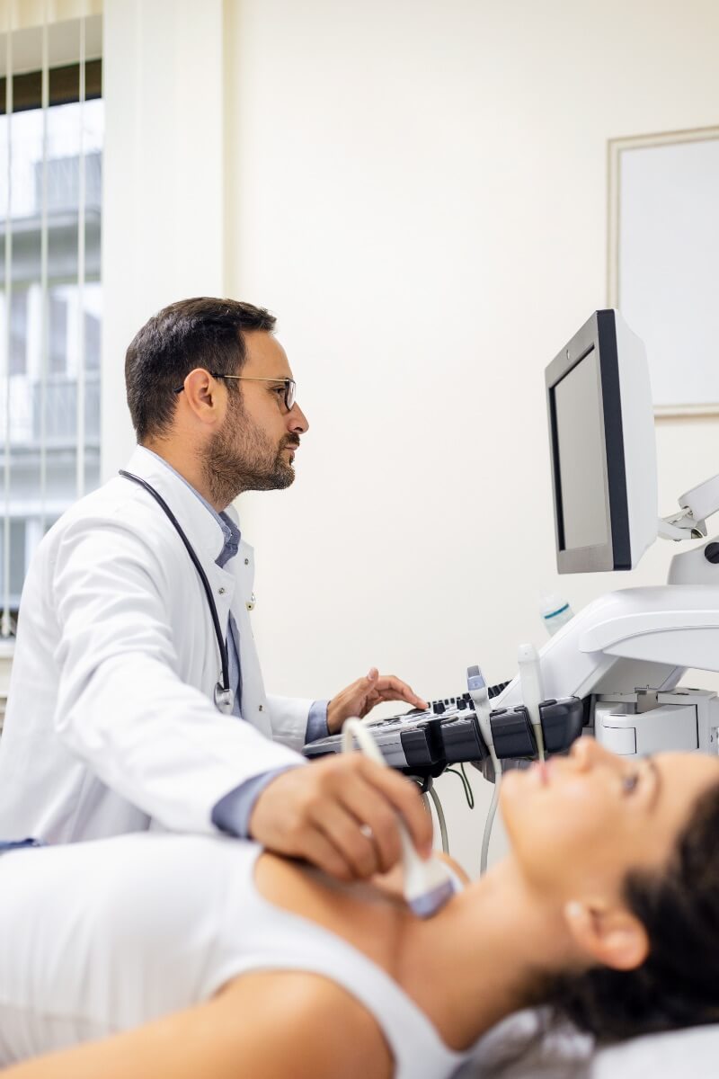
Ultrasound examinations
Ultrasound uses sound waves to detect heart and organ damage, swelling, infections, tumors, and other conditions. Ultrasound examinations can cover different parts of the body and organs, and some common ultrasound examinations include:
- Abdomen: Examination of abdominal organs such as the liver, gallbladder, spleen, pancreas, kidneys, and intestines.
- Pelvic Ultrasound: Examination of pelvic organs such as the uterus, ovaries, prostate, bladder, and intestines.
- Breast: Examination of the breasts to detect changes such as cysts or tumors.
- Thyroid: Examination of the thyroid gland to assess size, structure, and any changes.
- Heart (echocardiography): Examination of the heart to assess the function of the heart chambers, valves, and blood flow.
- Blood vessels (Doppler): Examination of blood vessels to assess blood flow, detect any narrowing or blockages.
- Musculoskeletal Ultrasound: Examination of muscles, tendons, ligaments, joints, and bones to detect injuries or conditions.
- Soft Tissues: Examination of soft tissues to detect changes, cysts, or tumors.
- Ultrasound of cervical lymph nodes – Carotid ultrasound: Examination of lymph nodes in the neck to assess any changes or swelling.
At Puls GO Diagnostic Center, we perform a range of ultrasound examinations, which you can read about on the Ultrasound page, while the prices for this type of diagnostic examination are available on this page.

CT Scans
A CT scanner provides a more detailed examination of your body compared to X-rays. CT scanning is a diagnostic procedure that uses X-rays and computer processing to obtain a detailed image of the interior of the body. The CT scanner uses a series of X-ray images taken from different angles to reconstruct a three-dimensional image of organs and tissues.
CT scanning can be performed for various parts of the body and purposes, including:
- Head CT: Examination of the brain to detect tumors, bleeding, injuries, or other conditions.
- Chest CT: Examination of the lungs, heart, blood vessels, or other structures in the chest to detect infections, tumors, embolisms, or other pathologies.
- CT of the Abdomen and Pelvis: Examination of the internal organs in the abdomen and pelvis, such as the liver, kidneys, intestines, uterus, ovaries, and prostate, to detect changes or diseases.
- CT of the Bones and Joints: Examination of bones, joints, and soft tissues to detect fractures, degenerative changes, tumors, or other injuries.
- CT Angiography: Examination of blood vessels to assess blood flow and detect narrowing, plaque, or aneurysms.
- CT Coronary Angiography: Examination of the heart and blood vessels.
- Ca Score – Calcium Score: Provides insight into the percentage of calcium deposits in blood vessels.
Before a CT scan, it may be necessary for the patient to receive contrast material intravenously to enhance the visualization of specific parts of the body.
It is important to note that each patient may have individual needs for CT scanning, and the decision regarding the type of scan and preparation is made by the doctor based on specific symptoms and the patient’s condition.
These are just some of the examinations that can be performed with a CT scanner. At Puls GO Diagnostic Center, we offer a range of examinations using this device. You can read more about them on our website, while the prices for CT scans are available on the Price List page.
X-ray imaging
X-ray imaging is a quick and simple diagnostic method. You lie or stand and position yourself according to the instructions. The imaging is completed in just a few minutes.
X-ray imaging is a diagnostic procedure that uses X-rays to obtain images of the internal structures of the body. This procedure is often used to detect bone fractures after accidents or injuries, but it can also be used to examine other parts of the body for the diagnosis of various conditions, such as respiratory problems, pneumonia, lung cancer, or other health issues.
During an X-ray imaging procedure, the patient is positioned between the X-ray machine and the detector, while X-rays pass through the body and create an image on the detector. The image is usually taken from multiple angles to provide a detailed view of the area of interest.

X-ray imaging can be performed for various parts of the body and purposes, including:
- Chest X-ray: Examination of the lungs to detect infections, inflammations, tumors, or other abnormalities.
- Bone and Joint X-ray: Examination of the bones to detect fractures, deformities, degenerative changes, or other injuries.
- Dental X-ray: Examination of the teeth to detect cavities, infections, inflammations, or other dental and jaw problems.
- Abdominal X-ray: Examination of the internal organs in the abdomen to detect changes, stones, obstructions, or other conditions.
- Spine X-ray: Examination of the spine for the diagnosis of issues such as herniated discs, deformities, or injuries.
During an X-ray imaging procedure, the patient will be asked to position themselves appropriately, and protective equipment may be used to reduce exposure of other body parts to radiation.
It is important to note that X-ray imaging uses X-rays, which are a form of ionizing radiation, so it is applied only when necessary and when the expected benefit outweighs the potential risks of radiation exposure. The doctor will determine the need for X-ray imaging and explain all the steps and any preparations required before the procedure.
To learn more about X-ray imaging at Puls Go Fast Diagnostic and Care Center, please read here, while the prices are available in our price list.
Magnetic Resonance Imaging (MRI)
MRI scanning. Instead of radiation, MRI uses radio waves and a magnetic field to create images of your body. This allows your radiologist to have a better view of soft tissue behind or inside your bones. It is particularly useful for imaging the brain and spinal cord, or for detecting torn ligaments or tumors. Inform your doctor if you are pregnant or have any metal or electronic implants, such as a pacemaker, artificial knee or other joints, dental fillings or bridges, or a cochlear implant for hearing. Your technician can adjust the procedure so that you can still undergo an MRI scan if you have any of these.
Pet scanner
PET (Positron Emission Tomography) scanning is a type of imaging used in the field of nuclear medicine. It involves the use of a small amount of radioactive material to examine the inside of your body at a molecular level. PET scans can detect cancer or problems with the heart, brain, nerves, and other parts of the body before other imaging methods can. You will typically need to refrain from eating or drinking, except for water, for several hours before the procedure.
What to expect during radiologist examination?
Depending on the procedure, your appointment may last only a few minutes or it can take 2 hours or more. Usually, you do not require any special preparation for the examination. However, some tests may require you to avoid certain foods, medications, and fluids before your arrival. Always inform our team if you are pregnant or trying to conceive. X-rays and CT scans involve low-dose radiation. Your doctor may prefer to use an alternative diagnostic test if possible to avoid exposing your baby to any potential risk.
Radiological treatments
Most radiologists assist in the diagnosis of health problems. However, some focus more on performing procedures and treatments.
Interventional radiologists introduce instruments through a small incision in your body where the treatment is needed. They may use:
- Needle for fluid drainage
- Tube for drainage or administration of medications or nutrition
- Laser for the removal of fibroids or other growths
- Stent, a small tube, to support a blood vessel
- Balloon for artery dilation (angioplasty)
Interventional radiologists sometimes take tissue samples for microscopic examination (biopsy) when cancer is suspected.
Radiation oncologists specialize in cancer treatment. They use energy beams or radioactive particles to target cancer cells while limiting damage to healthy cells. Your radiation oncologist will closely collaborate with all other members of the oncology treatment team.


