What is ultrasound?
Ultrasound, also known as sonography or simply ultrasound (US), is a medical imaging technique that uses high-frequency sound waves to visualize images of organs, tissues, and structures inside the body. Ultrasound examinations are non-invasive, painless, and do not involve the use of ionizing radiation, making them a safe and widely used diagnostic tool.
Ultrasound can help diagnose many conditions and diseases, such as tumors, cysts, inflammations, blood clots, and other changes in organs and tissues. It’s important to note that ultrasound is not always sufficient for diagnosing all conditions, but it can be used in combination with other diagnostic methods.
Who performs an ultrasound examination?
An ultrasound examination is usually performed by a radiology specialist, but it can also be performed by other specialists who are trained for ultrasound examination, such as specialists in gastroenterology, internal medicine, general medicine, etc.
How is the ultrasound examination performed?
During the ultrasound examination, a trained doctor will apply a special gel to the skin in the area being examined. This gel helps improve the transmission of sound waves and allows the ultrasonic probe, also known as transducer (eng. transducer), to move smoothly over the skin. The transducer emits high-frequency sound waves, which bounce off organs and tissues in the body, creating echoes that the transdcer registers and sends to a computer. The computer then uses these echoes to create detailed real-time images of the internal structures of the body.
Ultrasound examinations can be used to examine many parts of the body, including the stomach, pelvis, breast, thyroid gland, and musculoskeletal system, as well as to monitor fetal development during pregnancy. The specific preparation and procedure for ultrasound examination will depend on the part of the body being examined. Some ultrasound examinations may require the patient to fast for a certain period or to drink water before the examination to fill the bladder, which can help to get better images of the pelvic organs.
During the examination, the sonograph will move the probe across the skin, taking images from different angles to get a complete view of the area being examined. The patient may be asked to change position or hold their breath for a short time to obtain better images.
After the examination, the gel will be wiped off, and the patient will immediately be able to resume normal activities. The images obtained during the examination will be examined by a radiologist or other healthcare professional who will interpret the results and submit a report to the patient’s doctor.
Ultrasound examinations are a safe and effective diagnostic tool that can provide valuable information about the internal structures of the body without the need for invasive procedures or exposure to ionizing radiation.
Ultrasound of the abdomen
Abdominal ultrasound is used to visualize images of organs and structures in the abdominal cavity, including the liver, gallbladder, pancreas, spleen, kidneys and bladder. It is most used to diagnose and evaluate conditions such as:
- Gallstones or other gallbladder problems
- Liver disease or liver tumors
- Tumors or inflammation of the pancreas
- Kidney stones or other kidney problems
- Enlarged spleen or other spleen disorders.
- Abdominal aortic aneurysm
- Appendicitis
- Fluid accumulation in the abdominal cavity
How to prepare for ultrasound examination?
To prepare for an abdominal ultrasound, the patient will need to:
- Avoid consuming gas-producing foods (such as beans, cabbage, peas) a few days before the examination.
- Take gas-relief tablets one day before the examination.
- Fast for 5-6 hours prior to the examination, avoiding food and coffee.
- Drink 2-3 glasses of water one hour before the examination and refrain from urinating until the end of the examination.
The examination is performed in a supine position by the patient lying on his back, and in some situations the doctor may ask the patient to take a deep breath and hold his breath or turn to the left or right side.
At Puls GO diagnostic center, we perform ultrasound examinations that can cover various parts of the body and organs. Some common types of ultrasound examinations include:
Abdominal ultrasound
Abdominal ultrasound is a type of diagnostic test that uses high-frequency sound waves to create images of the organs in the abdomen. Ultrasound examination can help in diagnosing a wide range of conditions and diseases affecting the organs in the abdomen, including the liver, gallbladder, spleen, pancreas, kidneys, and abdominal aorta.
Ultrasound of the kidneys
Ultrasound of the kidneys by displaying pictures of the kidneys helps to diagnose and assess various conditions such as:
- Kidney stones
- Cysts or tumors of the kidneys
- Enlarged kidneys.
- Blockade in urine tract
- Congenital abnormalities
- Inflammation or infection in the kidneys
During the procedure, a trained doctor will apply a gel to the skin of the abdomen and use a probe to send sound waves through the kidney area.
Kidney ultrasound is often used in combination with other kidney function tests, such as blood tests or urine tests.
Ultrasound of the pancreas
The pancreas is a gland located in the upper part of the abdominal cavity, behind the stomach, and produces digestive enzymes and hormones, such as insulin.
Pancreatic ultrasound can help diagnose and evaluate various conditions, such as:
- Tumors or cysts of the pancreas
- Pancreatitis (inflammation of the pancreas)
- Blockage in the ducts of the pancreas
- Increased pancreas
- Collections of fluids around the pancreas
Pancreatic ultrasound is often used in combination with other tests, such as laboratory blood tests or CT scans.

Ultrasound of the breast
Breast ultrasound is used to evaluate lumps in the breast or other abnormalities detected on mammography or physical examination, as well as to conduct biopsy procedures.
Breast ultrasound can help:
- To distinguish between cysts (sacs filled with liquid) and solid masses in the breast,
- to provide information about the characteristics of any solid mass.
- To determine whether the mass is likely benign (non-cancerous) or malignant (carcinogenic).
It is important to note that breast ultrasound is not a substitute for mammography, which is still the primary tool for breast cancer screening in most women.
Ultrasound of the heart
What does a heart ultrasound show?
Ultrasound of the heart, also known as echocardiography, provides us with detailed information about the structure and function of the heart.
There are two main types of echocardiography: transthoracic echocardiography (TTE) and transesophageal echocardiography (TEE). TTE is the most common type of echocardiography, which is performed by placing a probe on the outside of the chest. TEE is performed by inserting a thin, flexible tube with a probe at its end into the esophagus to give a more detailed representation of the heart.
Ultrasound of the heart can help the cardiologist diagnose and evaluate various conditions and symptoms, such as:
- Disorders or abnormalities of the heart valves
- Congenital heart defects
- Enlarged heart or thickened heart muscle
- Heart failure
- Pericardial effusion (fluid around the heart)
- Blood clots or tumors in the heart
- Problems with the aorta or other blood vessels
- Pain in the chest
- Rapid heartbeat
- Skipping the heart
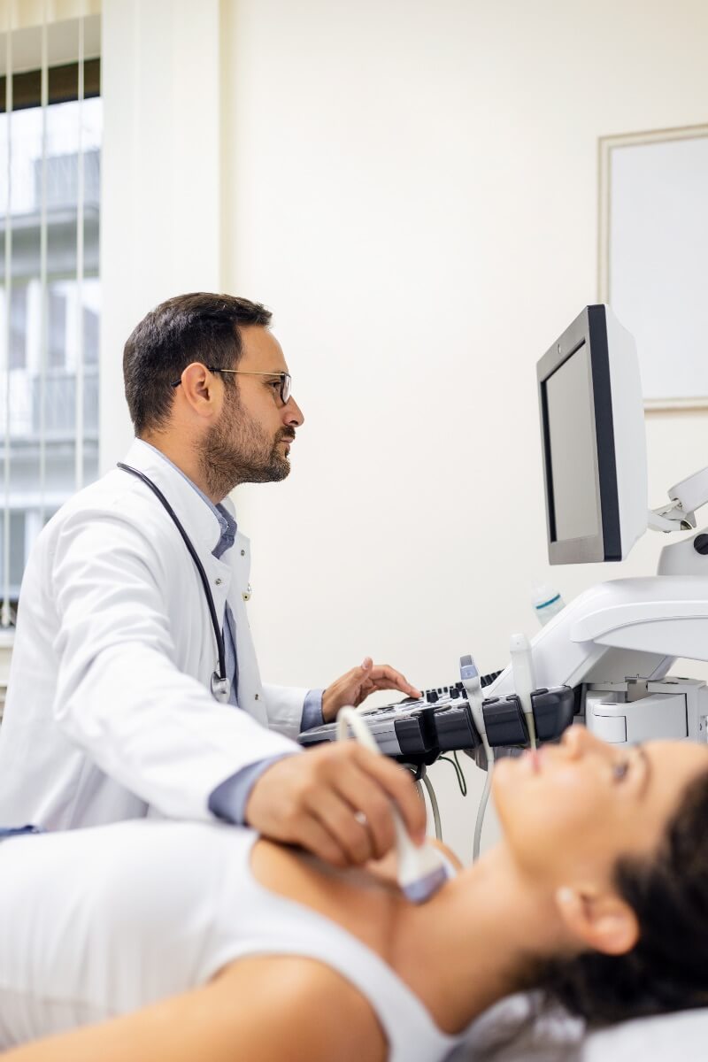
Ultrasound of the thyroid gland
Thyroid, or thyroid gland, is a gland with internal secretion of hormones located in the front of the neck and produces hormones that regulate metabolism. Ultrasound of the thyroid gland can help diagnose and evaluate various conditions, such as:
- Nodes or masses in the thyroid gland
- Enlargement of the thyroid gland (goiter)
- Thyroiditis (inflammation of the thyroid gland)
- Cysts or other fluid-filled structures in the thyroid gland
- Abnormal blood flow to the thyroid gland
Ultrasound of the thyroid gland can help distinguish between benign (noncancerous) and malignant (cancerous) thyroid nodes or masses, as well as provide information about the characteristics of any nodes or masses. This helps determine whether a biopsy is needed to further assess thyroid tissue.
It is often used in combination with other thyroid function tests, such as laboratory analyses or scintigraphy.

Ultrasound of the testicles
Testicular ultrasound helps in the diagnosis and evaluation of various conditions of the testicles and surrounding tissues, such as:
- Testicular torsion (twisted testicle)
- Trauma or testicular injury
- Tumors or masses of the testicles
- Varicocele (varicose veins in the scrotum)
- Hydrocele (fluid-filled bags around the testicles)
- Epididymitis (inflammation of the epididymis)
During the procedure, a trained doctor will apply the gel to the skin of the scrotum and use a probe to send sound waves through the area of the testicles.

When to see a doctor?
Ultrasound examinations are safe, effective diagnostic tools that allow visualization of the internal structures of the body without invasive procedures or exposure to ionizing radiation. They can help diagnose a variety of conditions and diseases, including tumors, cysts, inflammation, blood clots, and other changes in organs and tissues.
Keep in mind that it is crucial to consult your doctor if you are experiencing any symptoms, as he may provide accurate advice based on your individual medical history and current medical condition.
How much does an ultrasound cost?